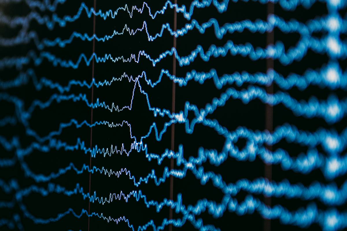
Electroencephalography (EEG) is a non-invasive method of studying brain activity that measures electrical signals on a large scale around the patient's skull. Spectrograms convert this raw EEG data into visual representations displaying frequency, time, and power intensity simultaneously, making trend analysis far more intuitive for clinicians.
During anesthesia induction with propofol, brain activity shifts to slower patterns. The spectrogram shows a brightening of power in the 8-12 Hz range combined with stronger low-frequency activity. This alpha-delta pattern indicates adequate cortical suppression and appropriate anesthetic depth.
Inhaled anesthetics like sevoflurane produce similar but distinct signatures, generating robust delta activity and less pronounced alpha power. Elderly patients may display weaker alpha oscillations despite adequate anesthesia, making spectrograms particularly valuable for individualized assessment in this population.
As anesthetic concentration increases, burst suppression may occur - alternating periods of high-amplitude activity and electrical silence. This pattern appears as dark bands on spectrograms and suggests profound cortical suppression, potentially foreshadowing delayed emergence.
Artifacts require careful interpretation. Electromyographic activity from facial muscles appears as high-frequency power, often forming a distinct band above 30 Hz. Clinicians must distinguish genuine anesthetic changes from electrode artifacts or neuromuscular blocker effects.
Spectrograms enable trend analysis over time, revealing subtle shifts that foreshadow clinical changes before vital signs reflect them, supporting safer and more personalized anesthetic management.